- Latest news▼
-
12:16, April 19 Scientists grow human mini-lungs in lab

-
10:23, April 19 JAMA Oncology: Urine test can help rule out high-grade prostate cancer with almost 100% accuracy, study shows

-
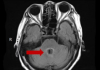
18:00, April 18 Daily Mail: Elderly woman in China gets infected with brain-eating amoeba
-
14:19, April 18 Obesity: exercising before breakfast helps you lose weight faster

-
10:42, April 18 The Conversation: childhood trauma can cause pathological hoarding

-
08:37, April 18 Daily Mail: Satiating food reduces cravings for sweets, nutritionist says

-
18:22, April 17 First Armenian-German Conference entitled “Heart Failure Spring School”

-
08:38, April 17 Why do kids usually recover from COVID-19 more easily than adults?
-
14:37, April 16 Daily Mail: intermittent fasting is not suitable for children and women before their periods

-
16:41, April 15 Cell: in carriers of defective BRCA2 gene, sugar consumption increases cancer risk

-
15:04, April 15 305 cases of measles recorded in Armenia so far in 2024

-
14:38, April 15 Food and Environmental Virology: tea contributes to effective coronavirus control

-
12:41, April 15 Daily Mail: vitamin A, B3 and E supplements can be dangerous

-
10:56, April 15 Diabetes Care: evening physical activity is good for the heart

-
08:27, April 15 Women are more susceptible to blood loss and death during bypass surgery than men, researchers say

All materials
3D bioprinted placenta model may lead to treatment for leading cause of maternal death
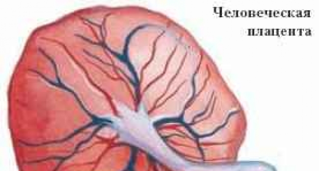
Despite preeclampsia being the leading cause of death from pregnancy there is a surprisingly little amount of information known about it, what causes it and how to prevent it. The condition occurs in about 5% of pregnancies worldwide, and while thankfully it isn’t always lethal, it does lead to approximately 30,000 deaths each year. Part of the problem with learning more about preeclampsia is the fact that the placenta is an organ that doesn’t really exist until a woman becomes pregnant, making it difficult to impossible to accurately study without putting the mother and fetus in jeopardy. Additionally, the human placenta is different from that in nearly all other mammals, so research and testing on an animal placenta wouldn’t lead to much useful data.
We may be on the road to understanding more about how the placenta works, and what happens when it doesn’t work, thanks to 3D bioprinting. A team of researchers brought together from from the Sheikh Zayed Institute for Pediatric Surgical Innovation at Children’s National Health System and the Tissue Engineering & Biomaterials Laboratory, Fischell Department of Bioengineering at the University of Maryland created the first 3D bioprinted placenta model and successfully used it in a preeclampsia study. Modern bioprinting has advanced to the level that the research team was able to successfully produce a placenta model that would accurately mimic the organ’s highly complex cellular structure. The placenta model provided the researchers with an opportunity to produce groundbreaking research that could lead to new, life-saving treatments for a wide range of pregnancy-related medical complications.
There are some medical theories that suggest that one of the causes of preeclampsia lies with a type of special cell in the placenta called trophoblasts. In a healthy placenta, the trophoblasts attach themselves to the uterine wall and then start to grow into the tissues of the uterus during the first stage of pregnancy, essentially bonding both organs together. At that point they connect to the mother’s blood vessels, which plays a vital role in helping to nourish and encourage growth in the fetus. The theory is that if the trophoblasts do not migrate normally it could lead to poor placental perfusion, the insufficient blood flow between a placenta and a uterus that causes preeclampsia.
In a new study that was published in American Chemical Society (ACS) Biomaterials Science & Engineering, the scientists detail their research which involved using the bioprinted model to observe the migration of trophoblasts cells form the placenta to the uterus. The bioprinted placenta model created by the research team contained key cellular, biochemical, and extracellular matrix components that accurately simulated the process of trophoblast migration. This is the first time scientists were able to successfully 3D bioprint a placenta and test cell migration of this type, and it could lead to several life-saving medical treatments.
“Our study provides a proof of concept that a 3D bioprinted placenta model is a viable way to study and understand the dynamics of cell migration in the formation of the placenta. What we have learned from this initial study is a significant step toward understanding the cause of preeclampsia and a potential therapy,” explained the chair of the Fischell Department of Bioengineering and the University of Maryland, John P. Fisher.
The team was able to successfully use the 3D bioprinted placenta model to track the results of introducing epidermal growth factor (EGF), a peptide that stimulates cell growth, proliferation, and differentiation, on trophoblast migration. The results strongly suggest that introducing the EGF to the placenta had a positive effect on the migration of the trophoblasts. While there is still more testing required, it seems that EGF may be a potential therapeutic treatment for preeclampsia. The reason that the 3D bioprinted model was able to provide such valuable data is the fact that the scientists were able to learn far more about the dynamic behavior of the trophoblast cell movements than more traditional 2D models.
When a 2D model is used researchers can only tell that a cell has moved, but with the 3D model they were able to see how it moved, where it moved and if a group of cells moved together. According to their research paper, the 3D bioprinted placenta model is a promising first step to developing a more sophisticated placenta model that could be bioengineered as a powerfultesting and research tool. Being able to accurately simulate the biological workings of an organ like a placenta could help them develop new treatments for preeclampsia and other similar placenta-related conditions including placenta accreta and placenta previa.
Follow NEWS.am Medicine on Facebook and Twitter

- Video


- Event calendar
- Archive
- Most read
month
week
day
- WHO: Nigeria pioneers revolutionary meningitis vaccine 1184
- One-third of women experience menstruation-related migraines, most often during premenopause - study 1171
- Daily Mail: vitamin A, B3 and E supplements can be dangerous 1024
- Food and Environmental Virology: tea contributes to effective coronavirus control 1016
- Cell: in carriers of defective BRCA2 gene, sugar consumption increases cancer risk 989
- 305 cases of measles recorded in Armenia so far in 2024 981
- Women are more susceptible to blood loss and death during bypass surgery than men, researchers say 969
- Diabetes Care: evening physical activity is good for the heart 925
- Daily Mail: intermittent fasting is not suitable for children and women before their periods 799
- First Armenian-German Conference entitled “Heart Failure Spring School” 580
- Why do kids usually recover from COVID-19 more easily than adults? 467
- Obesity: exercising before breakfast helps you lose weight faster 466
- The Conversation: childhood trauma can cause pathological hoarding 463
- Daily Mail: Elderly woman in China gets infected with brain-eating amoeba 443
- Daily Mail: Satiating food reduces cravings for sweets, nutritionist says 439
- Find us on Facebook
- Poll