- Latest news▼
-
12:16, April 19 Scientists grow human mini-lungs in lab

-
10:23, April 19 JAMA Oncology: Urine test can help rule out high-grade prostate cancer with almost 100% accuracy, study shows

-
18:00, April 18 Daily Mail: Elderly woman in China gets infected with brain-eating amoeba
-
14:19, April 18 Obesity: exercising before breakfast helps you lose weight faster

-
10:42, April 18 The Conversation: childhood trauma can cause pathological hoarding

-
08:37, April 18 Daily Mail: Satiating food reduces cravings for sweets, nutritionist says

-
18:22, April 17 First Armenian-German Conference entitled “Heart Failure Spring School”

-
08:38, April 17 Why do kids usually recover from COVID-19 more easily than adults?

-
14:37, April 16 Daily Mail: intermittent fasting is not suitable for children and women before their periods

-
16:41, April 15 Cell: in carriers of defective BRCA2 gene, sugar consumption increases cancer risk

-
15:04, April 15 305 cases of measles recorded in Armenia so far in 2024

-
14:38, April 15 Food and Environmental Virology: tea contributes to effective coronavirus control

-
12:41, April 15 Daily Mail: vitamin A, B3 and E supplements can be dangerous

-
10:56, April 15 Diabetes Care: evening physical activity is good for the heart

-
08:27, April 15 Women are more susceptible to blood loss and death during bypass surgery than men, researchers say

All materials
Nanotechnology sees upgrade with accelerated NanoMRI process
How do you view nanoscale materials, such as viruses and cells, up close and in-depth? You use a nanoMRI, of course. Now researchers from the Swiss Federal Institute of Technology in Zurich have accelerated the nanoMRI measurement process, bringing the technology one step closer to widespread commercial use.
Producing images of nano-sized objects with near-atomic resolution is hugely difficult. More importantly, the process takes time. A single nanoMRI scan requires not hours, not days, butweeks to complete.
In general, magnetic resonance imaging works by exploiting the fact that certain atoms have nuclei that act like tiny spinning magnets. If you bring these atoms into a magnetic field, they will rotate around the field’s axis the same way a spinning top rotates when it is imperfectly balanced and on a slant. This rotation is called precession and it happens at a precise frequency known as the Larmor frequency.
The Larmor frequency depends on both the type of atom and the general strength of the magnetic field. So to compose an image, an MRI essentially evaluates atoms’ locations by the frequencies at which they precess.
“When you look at a clinical MRI picture, you see bright pixels where the density of atoms is high, and dark pixels where the density is low,” Dr. Alexander Eichler, a postdoc researcher within the department of physics at the Swiss Federal Institute of Technology, stated in a press release.
When it comes to nanoMRI, though, the magnetic strength of nanomaterials is exceedingly small and so the sensitivity required of the technology is “at least a quadrillion times better,” Eichler explained. This has made it necessary to measure nano-sized information sequentially — one bit after another, a time-consuming process.
But what if you could measure the necessary information at the same time?
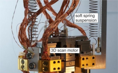
Such parallel measurement or multiplexing would require being able to distinguish where each bit of magnetic field information belongs in the final picture. While different strategies have been proposed, the research team in Zurich has developed a technique using a magnetic resonance force microscopy (MRFM) apparatus.
Here, the atomic nuclei experience a tiny magnetic force that is transferred to a cantilever, which acts as a mechanical detector. In response to the force, the cantilever vibrates and then, in turn, an image can be assembled from the measured vibration.
Using the new technique, a scan that normally might take two weeks can now occur within two days. Having overcome this major obstacle of high-resolution nanoMRI, the new research “brings us closer to the commercial implementation of nanoMRI,” Eichler concluded. In turn, this technology will allow scientists a closer view of cells and viruses, helping them advance their work on new medicines and vaccines.
Follow NEWS.am Medicine on Facebook and Twitter

- Video


- Event calendar
- Archive
- Most read
month
week
day
- WHO: Nigeria pioneers revolutionary meningitis vaccine 1176
- One-third of women experience menstruation-related migraines, most often during premenopause - study 1142
- Daily Mail: vitamin A, B3 and E supplements can be dangerous 967
- Food and Environmental Virology: tea contributes to effective coronavirus control 967
- Women are more susceptible to blood loss and death during bypass surgery than men, researchers say 958
- Cell: in carriers of defective BRCA2 gene, sugar consumption increases cancer risk 939
- 305 cases of measles recorded in Armenia so far in 2024 931
- Diabetes Care: evening physical activity is good for the heart 917
- Daily Mail: intermittent fasting is not suitable for children and women before their periods 749
- First Armenian-German Conference entitled “Heart Failure Spring School” 534
- Why do kids usually recover from COVID-19 more easily than adults? 412
- Obesity: exercising before breakfast helps you lose weight faster 404
- The Conversation: childhood trauma can cause pathological hoarding 395
- Daily Mail: Elderly woman in China gets infected with brain-eating amoeba 369
- Daily Mail: Satiating food reduces cravings for sweets, nutritionist says 367
- Find us on Facebook
- Poll