make preferable
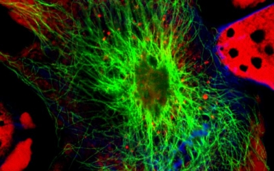
Silver-based nanoparticles are used with considerable success in the field of medicine. But the potential toxicity of their long-term use has always been a concern. The latest nanoparticle, however, invented by a group of scientists at the University of California Santa Barbara, leaves no question about its safety. Not only does it effectively penetrate and target tumor cells — due to its spherical shape and peptide casing — but excess particles that don't hit their target break down and get flushed away, reducing toxicity considerably, the Medical Daily reported.
The core of the nanoparticle employs a phenomenon called plasmonics, which reveals its electromagnetic properties. In plasmonics, laser radiation is used to resonate light off of metallic nanoparticles, like gold and silver. This causes so-called "plasmonic enhancements," which concentrate electromagnetic fields on the surface. When dyes are added, they appear far brighter than usual. When the core is etched, the enhancement goes away and the particle becomes dim.
What gives this method an advantage over the others is the etching process. Etching involves selectively removing the excess nanoparticles that do not hit their target. This method leaves only the intact nanoparticles for imaging or quantification, thus revealing which cells have been targeted and how much each cell internalized.
The intrinsic nature of nanoparticles such as their quantum properties and their ability to absorb and carry other compounds makes them useful in administering drugs that cannot penetrate cell membranes on their own, such as RNA and DNA genetic drugs. Besides this, silver is known to have several antibacterial properties, even against antibiotic-resistant organisms, and is effective in cancer treatments.