make preferable
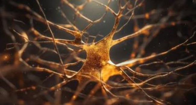
Using a 3D printer, researchers from Uppsala University have managed to create a model that resembles human nervous tissue. The sample, which can be grown from a patient's own cells, allows new drugs to be tested in the laboratory.
Motor neurons are nerve cells that control our muscles by sending signals from the brain and spinal cord to the body, Scientific Russia reported. In diseases such as amyotrophic lateral sclerosis (ALS), these cells are destroyed, leading to muscle weakness and paralysis.
On average, life expectancy after diagnosis is about 4 years, as the patient's ability to move and breathe gradually deteriorates. There is no cure for the disease yet, but some drugs can slow its progression.
In a new study published in the International Journal of Bioprinting, scientists have shown that it is now possible to use 3D printers to create models called organoids, which resemble human nervous tissue. Motor neuron organoids could be used in research or for testing new drugs, for example.
In the current study, scientists used human stem cells derived from skin and programmed to turn into motor neuron precursors, a type of immature nerve cells that can later develop into mature motor neurons. The cells were mixed with soft gelatin and then printed layer by layer on a 3D printer, creating the tissue and its structure. This three-dimensional distribution of cells in the biochannels improved their survival and nerve fiber growth.
In previous experiments, axons were only on the surface, but now the researchers were able to make them grow inside the biochar matrix as well. The solution to this problem was to use a softer 3D printing material (biochernyl) that retains its shape but still allows the axons to sprout in the material. To help the cells mature and develop, the scientists used small particles with a porous structure - mesoporous silica particles - that were infused with growth factors and added to the bioinks.
"For drug research and testing, it is important to be able to reproducibly print large numbers of organoids. Our method also allows us to incorporate other types of nerve cells, including glial cells, which will facilitate the creation of more complete models of the spinal cord," says Elena Kozlova.