make preferable
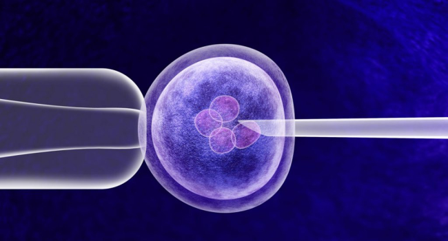
Researchers in China have identified critical mechanisms behind the developmental failure of many human embryos during the earliest stages of growth, offering potential new strategies to improve outcomes in in vitro fertilization (IVF), Xinhua reported. The findings were published in the journal Cell.
The study was led by scientists from Tsinghua University, who produced the first high-resolution recordings of human embryos during the first five days after fertilization.
Embryo developmental arrest remains a major challenge in IVF treatment. More than half of fertilized human eggs fail to reach the blastocyst stage, when embryos become suitable for implantation into the uterus. The biological reasons behind this high failure rate have remained largely unclear.
To investigate the problem, the researchers developed a specialized dual-view light-sheet microscope capable of continuously imaging living embryos with high clarity while minimizing damage to the delicate tissues.
Using this technology, the team analyzed more than 2,000 cell divisions in over 150 human and non-human primate embryos. Their results showed that more than 70% of embryos that stopped developing experienced abnormalities during the second cell division.
The researchers found that defects in the spindle—a cellular structure responsible for ensuring the accurate distribution of chromosomes during cell division—were a major cause of developmental failure. Abnormal spindle formation led to chromosome segregation errors, which subsequently triggered developmental arrest within a few additional cell cycles.
The study also identified a crucial role for centrosomes, cellular organelles involved in spindle assembly. When embryos contained an abnormal number of centrosomes, spindle function was disrupted, increasing the likelihood of chromosome errors.
Based on these observations, the scientists tested whether correcting centrosome-related defects could improve embryo development. They treated embryos with a low dose of a drug that regulates centrosome replication during the second cell division.
The intervention produced promising results: the proportion of embryos with a normal centrosome count increased from 40% to 80%. Importantly, the treatment did not affect embryos that already had normal centrosomes.
According to Chun So, the findings could eventually help reduce the incidence of early embryo developmental arrest in IVF clinics.
The research team plans to continue developing advanced imaging technologies to study later stages of human embryonic development after implantation, with the ultimate goal of improving pregnancy success rates and advancing reproductive medicine.