make preferable
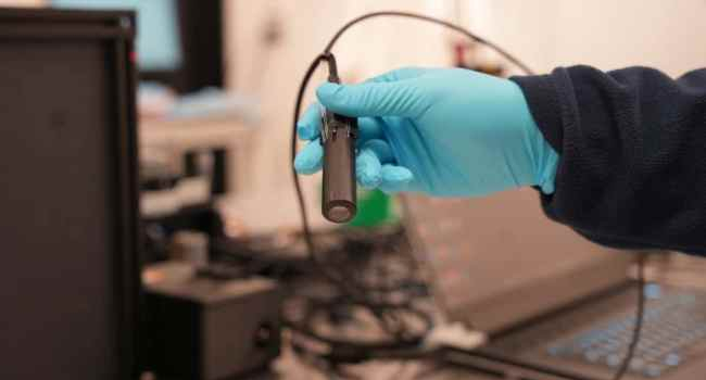
Researchers at Rice University have developed PrecisionView, a compact artificial intelligence (AI)-powered handheld endomicroscope designed to improve the early detection of epithelial cancers while offering an affordable alternative to existing diagnostic technologies.
Epithelial cancers account for approximately 80–90% of all cancer cases worldwide. Because these malignancies originate in the tissues lining organs, internal passageways, and the skin, early diagnosis is essential for improving survival. However, current diagnostic approaches often rely on invasive tissue biopsies.
Scientists believe that simultaneously examining cellular structures and the surrounding microvasculature can significantly improve the identification of cancerous and precancerous changes, helping physicians make more informed decisions about whether a biopsy is necessary. Although in vivo microscopy has shown promise for this purpose, current systems are limited by image quality, spatial resolution, and a relatively small field of view.
To address these challenges, the Rice360 Institute for Global Health Technologies developed PrecisionView, a portable device that combines AI with dual-modality fluorescence and reflectance imaging. The system provides real-time visualization of cellular structures and blood vessels over much larger tissue areas than conventional endomicroscopes. Compared with currently available clinical systems, PrecisionView delivers a fivefold larger field of view and an eightfold improvement in spatial resolution while maintaining high-quality cellular imaging.
The proof-of-concept prototype, described in the Proceedings of the National Academy of Sciences, is intended for imaging superficial epithelial tissues and was specifically designed to remain inexpensive, making advanced diagnostic imaging more accessible in low-resource healthcare settings.
The device incorporates commercially available miniature optical components together with deep learning algorithms that optimize image acquisition and reconstruction. It captures images at approximately 15 frames per second and automatically stitches multiple image tiles into detailed maps showing both cell nuclei and microvascular structures.
To evaluate the technology, researchers led by Rebecca Richards-Kortum and Ashok Veeraraghavan, together with collaborators from the University of Texas MD Anderson Cancer Center, tested PrecisionView using tissue samples from porcine tongue, human breast tissue, and cervical specimens containing precancerous lesions.
The resulting high-resolution images clearly identified important anatomical features, including transitions between different epithelial cell types. The system successfully distinguished abnormal tissue from healthy surrounding tissue, suggesting that it could help clinicians determine when a biopsy is warranted.
The team also performed real-time imaging inside the oral cavities of healthy volunteers. Thanks to the device's extended depth of field and wide imaging area, researchers obtained detailed images of cellular and vascular structures without repeated refocusing. The overlapping image frames were seamlessly combined into large-scale maps of the oral mucosa, revealing distinct microscopic features.
The Rice360 Institute focuses on creating affordable medical technologies for underserved regions, and PrecisionView was developed in response to the global shortage of accessible, high-quality diagnostic tools.
According to first author Huayu Hou, many healthcare facilities—particularly those with limited pathology services—lack timely access to the cellular-level information needed to guide diagnosis and treatment. Existing endomicroscopy systems often remain too expensive, technically complex, and limited in imaging capability for widespread point-of-care use.
Hou noted that the first functional prototype was completed in approximately one year, and the research team is continuing to improve both image quality and ease of use. Current efforts are focused on providing even higher-resolution imaging of living tissues in real time while simplifying the device for routine clinical practice.
Researchers are also developing AI-based diagnostic algorithms capable of automatically analyzing both cellular and vascular features to provide clinicians with real-time decision support. According to co-first author Jimin Wu, such capabilities could be especially valuable in regions where access to pathology specialists is limited.
Clinical validation studies are already underway. One trial is evaluating PrecisionView for head and neck cancer detection in collaboration with MD Anderson Cancer Center, while another planned study in Mozambique will assess the device's performance for cervical cancer screening in low-resource healthcare settings.