- Latest news▼
-
19:41, April 25 Children’s Hospital Los Angeles and International Center of Professional Development Allergy/Immunology Conference

-
17:31, April 25 JAMA: patient grew long, curly eyelashes because of chemotherapy
-
11:08, April 25 Mpox epidemic declared in Republic of the Congo

-
08:31, April 25 OU: quitting smoking 4 times more likely to cure laryngeal cancer

-
01:20, April 25 Paralyzed man in China writes hieroglyphs using neural implants placed in his brain

-
15:11, April 24 Zombie deer disease possibly linked to hunters’ deaths

-
12:27, April 23 Appetite: Scientists found out the secret to the appeal of large portions of fast food

-
10:33, April 23 Scientists test new approach to fighting viruses

-
08:38, April 23 Ketamine may help with postpartum depression

-
22:12, April 22 Unhealthy amount of sugar found in baby food products of a well-known brand

-
19:41, April 22 Air pollution puts health of more than 1.6 billion workers globally at risk

-
17:25, April 22 Scientists found baked goods and lack of sleep to be more dangerous than alcohol

-
16:02, April 22 342 cases of measles recorded in Armenia so far in 2024

-
15:29, April 22 BrainStimulation: electrical brain stimulation alleviates anxiety and depression in the elderly

-
08:27, April 22 Cognitively stimulating jobs in midlife could lower dementia risk in old age, study finds

All materials
How you can get the best, most accurate mammogram
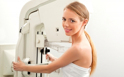
You probably never thought you could have a role in ensuring that your mammogram is as accurate as possible in detecting breast cancer. Women often don’t realize they can improve the quality of their mammography results by doing a few simple, critical things:
1. Cooperate as the technologist positions you
A screening mammogram consists of two views of each breast: one view in which the breast is compressed from top to bottom (called craniocaudal) and one view in which the breast is compressed at a 45 degree angle (called mediolateral oblique).
The technologist’s goal is to give every patient the best mammogram. In order to detect breast cancer, the radiologists must see the entire breast. The technologist attempts to pull as much breast tissue into the picture as possible.
While it may not be the most comfortable positioning, the technologist is working to include all the breast tissue, especially the extreme posterior breast tissue, in order to be sure that breast cancer is not missed simply because it was not included on the image.
Work with your technologist by being as cooperative and accommodating as you can while he or she positions you. Remember that the technologist must make sure your entire breast is included on the image for the best result.
2. Allow for breast compression
Mammography compresses the breast tissue. Normal breast tissue flattens like a sponge with compression. Breast masses and especially breast cancer do not flatten with compression and actually become more conspicuous, which enables the radiologist to detect them.
Allowing for adequate compression will enable the radiologist to see through the breast tissue and better detect breast cancer. Remember that while compression is uncomfortable, it’s only necessary for a few moments. The more you can tolerate with an attitude of patience, the more you are supporting the most accurate reading.
3. Try not to move; hold your breath while images are taken
When a patient moves during the exam, it limits the evaluation of the breast tissue. In order to read a mammogram, the radiologist enlarges each mammographic image on the viewing station. The smallest amount of motion during the examination can result in blurring of the image.
Calcifications, which can be an early sign of breast cancer, are not easily detected when the mammographic image is blurred by motion. Therefore, it is very important to hold your breath when the technologist is taking the image.
4. Bring prior mammogram images with you for comparison
When reading a mammogram, the radiologist compares it to prior images, which are essential to helping him or her detect small changes from year to year. Giving the radiologist reference to past exams gives you the best chance of early, subtle breast cancer detection. Therefore, if you have prior mammograms, be sure to bring the images to your radiologist.
Follow NEWS.am Medicine on Facebook and Twitter

- Related News
- Ketamine may help with postpartum depression In the end, only 6.7 per cent of the mothers who took S-ketamine experienced a major depressive episode...
- Daily Mail: vitamin A, B3 and E supplements can be dangerous And some supplements affect the effectiveness of medicines...
- Diets are pointless in case of acne, dermatologist says They invent different things, as if you can't eat dairy products, eggs, chocolate…
- Chinese tai chi helps lower blood pressure, study finds The traditional, slow-moving form of Chinese martial art helps better than aerobic exercises…
- Measure blood pressure 2 times a day: Armenia cardiologist gives advice on weather fluctuations The number of people applying to hospitals with high blood pressure and exacerbation of other heart diseases has increased…
- Video


- Event calendar
- Children’s Hospital Los Angeles and International Center of Professional Development Allergy/Immunology Conference
- First Armenian-German Conference entitled “Heart Failure Spring School”
- Allogeneic bone marrow transplant in case of hematological malignancy performed in Armenia for first time
All materials
- Archive
- Most read
month
week
day
- JAMA Oncology: Urine test can help rule out high-grade prostate cancer with almost 100% accuracy, study shows 1270
- Scientists grow human mini-lungs in lab 1156
- Next pandemic likely to be triggered by flu - scientists 917
- Scientists found baked goods and lack of sleep to be more dangerous than alcohol 837
- 342 cases of measles recorded in Armenia so far in 2024 795
- Scientists develop new method to safely stimulate immune cells to fight cancer 763
- Blood test can determine who is at risk of developing multiple sclerosis - scientists 762
- Cognitively stimulating jobs in midlife could lower dementia risk in old age, study finds 757
- BrainStimulation: electrical brain stimulation alleviates anxiety and depression in the elderly 701
- Unhealthy amount of sugar found in baby food products of a well-known brand 576
- Air pollution puts health of more than 1.6 billion workers globally at risk 572
- Ketamine may help with postpartum depression 559
- Appetite: Scientists found out the secret to the appeal of large portions of fast food 558
- Scientists test new approach to fighting viruses 541
- Zombie deer disease possibly linked to hunters’ deaths 430
- Find us on Facebook
- Poll