make preferable
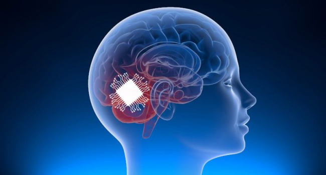
Bioengineering researchers at the Harvard John A. Paulson School of Engineering and Applied Sciences (SEAS) have developed a soft, thin, stretchable bioelectronic device that can be implanted into a tadpole embryo’s neural plate, the early-stage, flat structure that folds to become the 3D brain and spinal cord. The work was published in the journal Nature.
“Autism, bipolar disorder, schizophrenia – these all could happen at early developmental stages,” said Jia Liu, Assistant Professor of Bioengineering at SEAS. “There is just no ability currently to measure neural activity during early neural development. Our technology will really enable an uncharted area.”
The team of researchers developed a bioelectronic device based on fluorinated elastomers, which are comparable in softness to living tissue, but are also durable and can contain multiple sensors. These materials have made it possible to create implants that can “grow” into the brain, following its natural development, and track activity at all embryonic stages, from the flat neural plate to the formation of the 3D structure of the brain.
Previously, similar technologies have been tested on organoids—mini-organs made from stem cells in a Petri dish. However, working with live amphibian embryos was more difficult, as tadpole tissue was softer than artificial cultures, and a complete redesign of the materials and structure of the device was required.
“If we can fully leverage the natural development process, we will have the ability to implant a lot of sensors across the 3D brain noninvasively, and at the same time, monitor how brain activity gradually evolves over time,” Liu said. “No one has ever done this before.”
The work is an important step towards new approaches to brain research, early diagnosis of neuropsychiatric disorders, and the creation of the next generation of high-precision brain-machine interfaces.