make preferable
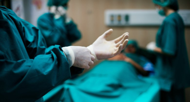
Researchers at Johns Hopkins University have developed an artificial intelligence model that can detect previously hidden signals in standard electrocardiograms (ECG) to accurately predict life-threatening complications after surgery. The technology significantly outperforms existing risk assessment methods currently used by physicians. The study was published in the *British Journal of Anaesthesia (BJA).
“We showed that even a basic ECG contains critical information that cannot be recognized by the human eye. Only machine learning methods can extract it,” said lead author Robert D. Stevens, head of the Division of Informatics, Integration and Innovation.
After major surgical interventions, a significant proportion of patients face severe complications — heart attack, stroke, or death within 30 days. Current risk scales predict such outcomes with only about 60% accuracy.
The researchers hypothesized that ECGs may contain hidden markers not only linked to heart health, but also to inflammation, metabolism, hormonal regulation, and electrolyte balance. To test this, they analyzed preoperative ECG data from 37,000 patients who underwent surgery in Boston.
Two algorithms were trained: one based solely on ECGs, and a second “fusion model” that combined ECG data with clinical information such as age, sex, and comorbidities.
Both outperformed existing risk scales, but the fusion model proved most effective, predicting complications with 85% accuracy.
“It’s remarkable that just 10 seconds of ECG data can so accurately forecast surgical outcomes. This is a truly meaningful result that could transform the way we assess surgical risk,” said study co-author Carl Harris, a PhD student in biomedical engineering.
The next step will be to validate the algorithm on even larger datasets and test it in real-time among patients preparing for surgery.