make preferable
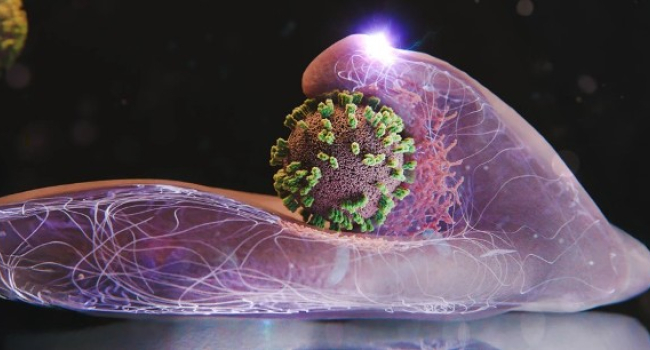
For the first time, scientists have managed to observe—in real time and with extremely high resolution—how influenza viruses enter living cells. This breakthrough became possible thanks to a new microscopy technology that could significantly improve the development of antiviral drugs.
The study was published in PNAS.
As winter arrives, so do the familiar symptoms: fever, body aches, and congestion. All are caused by influenza viruses, which enter the body through tiny droplets and then infect cells.
Researchers from Switzerland and Japan have now examined what was previously impossible to see. Using a new method they developed, they were able to approach the surface of living human cells in a Petri dish closely enough to observe the process of viral entry in real time and in remarkable detail.
The team led by Professor Yohei Yamauchi, a molecular medicine specialist at ETH Zurich, was particularly surprised by one finding: cells are not passive at all. They do not simply endure the virus’s intrusion — they actively attempt to “capture” it. Professor Yamauchi compares this to a dance — a complex interaction in which both sides move and respond to each other.
Although infection provides no benefit to the cell, the dynamic arises because the virus exploits a mechanism the cell uses every day. This process allows the cell to absorb hormones, cholesterol, iron, and other vital substances. For these substances — and for the virus — to enter the cell, they must bind to specific receptor molecules on the cell surface.
The virus’s behavior resembles surfing along the membrane. It “glides,” attaching first to one molecule, then another, until it finds a receptor-rich area — the perfect entry point. Once the receptors “realize” a virus has attached, an indentation begins to form. This indentation is stabilized by a protein called clathrin, which creates a structural framework. The indentation becomes a pocket, then a vesicle that is drawn inside the cell. Its membrane dissolves, and the virus is released into the cell.
Until now, such processes could only be studied using techniques that destroy cells, such as electron microscopy, which provides only static images. Another common approach, fluorescence microscopy, cannot reveal extremely fine details.
The new technology combines atomic force microscopy with fluorescence imaging. Named ViViD-AFM, it was developed specifically for studying viruses. It allowed the researchers to capture the entire infection process, almost like frame-by-frame footage. They showed that the cell actively assists the virus: it directs more clathrin to the point of entry and even lifts its membrane slightly to “push” the virus inward. If the virus tries to move away, these wave-like motions intensify.
This technology opens new possibilities for antiviral drug development. Scientists can now observe how potential treatments act on cells at the very moment of infection. The authors note that the method may also prove useful for studying other viruses, as well as vaccines.