make preferable
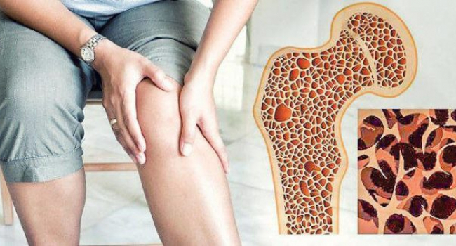
Researchers at the University of Texas at San Antonio have developed a biomaterial capable of simultaneously stimulating the growth of bone tissue and blood vessels in severe injuries involving substantial tissue loss. The study was published in the journal Biomaterials Advances.
One of the major challenges in treating large bone and vascular defects is that cells located in the center of the wound often receive insufficient blood supply and die. This can lead to graft failure and long-term complications.
To address this problem, the researchers created a three-dimensional scaffold made from two naturally occurring proteins in the body: collagen and fibrin. The material consists of interpenetrating polymer networks in which the proteins are intertwined, forming a supportive environment for tissue regeneration.
Essentially, the structure functions as a biological “construction framework,” providing cells with both the space and the biochemical signals needed to rebuild damaged tissues.
The scientists incorporated two types of cells into the hydrogel: microvascular fragments (MVFs), which can generate new blood vessels, and mesenchymal stem cells (MSCs), which can differentiate into bone-forming cells under appropriate conditions.
Importantly, these cells were distributed throughout the entire volume of the biomaterial rather than being confined to its surface, a feature considered critical for the successful repair of large tissue defects.
A key outcome of the study was the identification of the optimal ratio of the scaffold’s components. After testing five different formulations, the researchers found that a 50:50 ratio of collagen to fibrin provided the best balance between blood vessel formation and bone regeneration.
Fibrin was particularly effective at promoting vascular growth but created a less stable environment for bone development. Collagen, on the other hand, offered structural strength but tended to limit blood vessel infiltration.
By combining the two proteins in equal proportions, the team was able to support both processes simultaneously. Blood vessels formed extensive branching networks, while bone-forming cells successfully activated the genes required for generating new tissue.
The authors note that the use of natural components—including collagen, fibrin, and potentially a patient’s own cells—could reduce barriers to future clinical applications.
If further studies confirm its effectiveness, this approach may offer a promising strategy for treating complex injuries, large bone defects, and other conditions requiring coordinated regeneration of both skeletal tissue and blood vessels.