make preferable
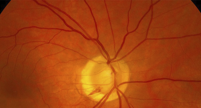
A standard digital photograph of the fundus can accurately predict the risk of heart attack or stroke within the next 10 years. This conclusion was reached by scientists from the University of Dundee (Scotland). The results of the study were published in the journal Cardiovascular Diabetology (CD).
The fundus is the inner surface of the eye opposite the lens, which includes the retina and the optic disc. Blood vessels pass through the fundus, supplying the retina with nutrients. The optic nerve, which transmits information to the brain, is also located here.
Any changes or damage to these structures not only negatively affect vision quality but can also be signs of other pathologies. With this in mind, scientists have developed an AI-based algorithm that can assess individual cardiovascular risk in a fraction of a second. The tool analyzes changes in the retinal vessels — such as narrowing, blockage, or damage — and correlates them with the risk of heart attack or stroke.
The development has already been tested on 1,200 volunteers. Those who showed the greatest increase in risk between two examinations three years apart were 54% more likely to develop heart and vascular disease.
The accuracy of the prediction based on a single image reached 70%. When data on classic risk factors and genetic predisposition were added, the accuracy rose to 73%. The authors believe that such technologies can be implemented in routine practice, for example, during visits to the ophthalmologist.
“This will help identify people at risk in advance and take preventive measures in a timely manner, from lifestyle changes to prescribing medication,” the researchers explained.