make preferable
A new study using advanced neuroimaging techniques has shown that SARS-CoV-2 infection can cause lasting changes in the brain, even in individuals who feel fully recovered. The research identified specific alterations in tissue microstructure and chemical levels that distinguish people with long COVID from those who recovered without residual symptoms, as well as from individuals who were never infected. The results were published in the journal Brain, Behavior, & Immunity – Health.
The study included 47 participants from the Gold Coast region in Queensland, Australia. The sample consisted of 19 people diagnosed with long COVID according to World Health Organization guidelines, 12 people who had recovered from COVID-19 without lingering symptoms, and a control group of 16 healthy individuals with no history of SARS-CoV-2 infection. All participants were aged 18 to 65.
Researchers excluded participants with other serious medical conditions to ensure results were specific to COVID-19. Participants completed detailed questionnaires assessing symptoms, including pain levels, physical function, fatigue severity, and cognitive impairment. Those with long COVID reported significantly higher levels of pain and fatigue compared to other groups, along with lower physical and cognitive performance scores.
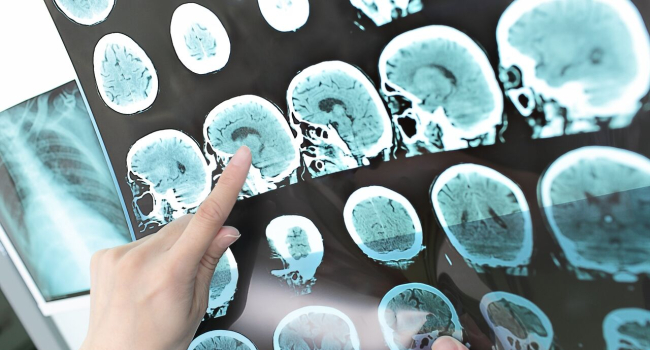
Participants underwent scanning with a 3-Tesla MRI. Three specific types of data were collected. First, T1-weighted and T2-weighted imaging was used to generate ratio maps, allowing researchers to estimate myelin content in the brain. Myelin is the protective sheath around nerve fibers that facilitates rapid communication between neurons.
Second, the team used diffusion-weighted imaging, which tracks the movement of water molecules in brain tissue, revealing microscopic architecture and the integrity of white matter pathways. Finally, magnetic resonance spectroscopy was applied, acting like a virtual biopsy to measure levels of specific chemicals in targeted brain regions. The chemical analysis focused on the posterior cingulate cortex, a center involved in memory and emotion.
The study found significant differences in T1w/T2w ratio maps, a marker of myelin signal intensity. Comparing long COVID patients to healthy controls revealed increased signal intensity in the precentral gyrus and middle temporal gyrus. The precentral gyrus is important for motor control, while the middle temporal gyrus supports memory. This increase may indicate remyelination or inflammation in these areas.
Comparisons between recovered individuals and long COVID patients revealed even more pronounced differences. Recovered participants showed higher signal intensity in the brainstem and cerebellum compared to long COVID patients. The brainstem controls vital functions such as sleep-wake cycles and pain processing, while the cerebellum coordinates voluntary movements and balance. Lower signal intensity in these regions in long COVID patients may relate to persistent fatigue and physical limitations.
Notably, even fully recovered individuals showed changes. Compared to the never-infected control group, recovered participants exhibited higher signal intensity in the precentral gyrus and posterior cingulate cortex, suggesting that the virus can leave a mark on the brain even without overt symptoms, possibly reflecting compensatory changes post-infection.
Diffusion-weighted MRI provided additional evidence of structural alterations. Long COVID patients showed lower mean diffusivity in the dentate regions of the cerebellum compared to healthy controls, indicating possible restriction of water movement due to edema or tissue remodeling. Recovered participants also had lower diffusivity in the caudate nucleus, a structure involved in motor control and learning, compared to controls.
Magnetic resonance spectroscopy revealed significant metabolic imbalances, mainly between the long COVID group and recovered participants. Long COVID patients had markedly reduced glutamine levels, an amino acid important for immune function and energy production, suggesting depletion due to ongoing inflammation or immune dysregulation. Conversely, long COVID patients exhibited higher N-acetyl-aspartate (NAA) levels compared to recovered participants. NAA is a marker of neuronal health and metabolism; higher levels in this context may indicate a compensatory response, with the brain working harder to maintain function under metabolic stress.
The researchers also analyzed correlations between these biological markers and participants’ symptoms. In long COVID patients, lower myelin signal in the middle temporal gyrus correlated with worse physical function, while signal intensity in the brainstem negatively correlated with cognitive performance. These relationships provide a biological basis for the subjective symptoms reported by patients.
“This study clearly shows that even individuals with no symptoms after recovering from COVID-19 can experience long-term effects of the virus on the brain,” said Tapalia.
However, the study has limitations. It was cross-sectional, capturing a single time point rather than longitudinal changes, so it cannot determine whether the observed brain alterations are permanent or evolve over time. The sample size was relatively small (fewer than 50 participants), which may limit reproducibility in larger populations. Statistical methods used to identify brain clusters involved thresholds that could introduce errors, though standard corrections were applied. The research is exploratory, aimed at generating hypotheses rather than providing definitive clinical diagnostic tools.
Further studies are needed to confirm these findings in larger, more diverse populations. Longitudinal studies tracking individuals from infection through recovery or development of long COVID would help determine whether brain changes resolve over time and clarify the timing of remyelination and metabolic adjustments.