make preferable

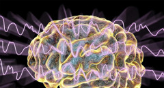
Panic disorder is associated with persistent, though moderate, changes in brain structure. This conclusion was reached by an international team of researchers who analyzed nearly 5,000 MRI scans from individuals with this diagnosis and from people without mental disorders. The study was published in the journal Molecular Psychiatry (MP).
Panic disorder (PD) is characterized by recurrent episodes of intense fear accompanied by rapid heartbeat, shortness of breath, dizziness, a sense of losing control, and other pronounced physical symptoms. According to experts, about 2–3% of the population experience PD at some point in their lives.
Until now, neurobiological studies of panic disorder have often relied on small sample sizes, making results difficult to reproduce. To overcome this limitation, scientists combined data within the framework of the international ENIGMA consortium.
“Neuroanatomical studies of panic disorder typically include small numbers of participants, which often leads to inconsistent findings,” explained the study’s authors, Laura Hahn and Mojtaba Agahjanyan. According to them, the project’s goal was “to conduct the most robust test to date of whether panic disorder is associated with persistent changes in brain structure.”
As part of a so-called mega-analysis, the researchers examined MRI data from nearly 5,000 individuals aged 10 to 66, including about 1,100 patients with panic disorder and 3,800 healthy volunteers. Standardized protocols and the FreeSurfer software package were used to accurately measure cortical thickness, surface area, and the volume of subcortical structures.
The results showed that people with panic disorder had, on average, slightly thinner cerebral cortex and smaller surface areas in the frontal, temporal, and parietal regions compared to healthy participants. In addition, reduced volumes of the thalamus and caudate nucleus were identified — structures involved in processing emotionally significant information and regulating stress responses.
“These regions play a key role in the perception, processing, and control of emotions,” the authors noted. The analysis also indicated that some changes depend on age, and that early onset of panic disorder (before age 21) is associated with enlarged lateral ventricles of the brain.
According to the researchers, the findings support contemporary models of panic disorder as a disruption of neural networks responsible for emotional regulation. In the future, this may help improve risk assessment, predict the course of the condition, and guide treatment selection.
The authors emphasized that the identified differences are moderate and cannot be used for diagnosis at the individual level. Nevertheless, the large-scale approach opens the way for more detailed “mapping” of mental disorders and comparison of their neurobiological foundations.
Follow NEWS.am Medicine on Facebook and Twitter