make preferable
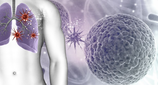
A newly developed artificial intelligence system may improve early detection of lung cancer by analyzing CT scans in a more advanced way. The model uses a dual-scale approach, allowing it to examine both detailed structures and the overall anatomical context at the same time—similar to how clinicians interpret medical images, but without needing to switch between views.
Researchers trained the system on CT scans from both healthy individuals and patients with lung cancer. This enabled the AI to distinguish between normal tissue, benign abnormalities, and malignant tumors.
The method could be especially useful for identifying lung cancer at an early stage, when tumors are small and difficult to detect. The model demonstrated an accuracy exceeding 96%, outperforming existing techniques while maintaining consistent results across different tests.
Early diagnosis is critical in lung cancer, as survival rates can rise dramatically—from around 10% in advanced stages to over 90% when the disease is detected early. However, interpreting CT scans remains challenging due to subtle tumor features and the possibility of human error.
To address this, researchers at Kaunas University of Technology developed an AI system that integrates both fine-detail analysis and a broader view of the lungs into a single process. This eliminates the need for radiologists to manually switch between perspectives, potentially saving time and reducing the risk of missed findings.
The system can evaluate small nodules while also understanding their location and importance within the entire lung structure. Experts describe this as having both a “zoomed-in” and “full-picture” view simultaneously.
Despite promising results, the technology still requires further validation before clinical use. Scientists emphasize the need for larger and more diverse datasets, as well as real-world testing.
If confirmed, this AI tool could support doctors by improving diagnostic accuracy, speeding up scan analysis, and reducing false positives. Rather than replacing clinicians, it would serve as a decision-support tool to help flag suspicious cases.
Researchers also believe similar AI techniques could be applied to other areas of medical imaging, including brain tumors, breast cancer, and eye diseases.