make preferable
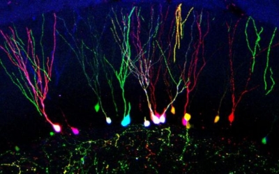
Scientists have developed a way of color-marking individual brain cells that promises to help researchers better understand the workings of the brain. In the journal Scientific Reports, researchers led by the University of Southampton in the UK describe how they developed and tested the technique in cell cultures and mice.
The human brain is a complex organ comprising billions of cells. The so called multicolor RGB tracking technique allows scientists to encode individual cells with unique heritable color markers. Once a cell is encoded, the unique color marker is embedded permanently in its DNA and also passes on to its daughter cells.
The aim of this new technique is to help neuroscientists see what is happening at the cell level.
The technique has been proved the effective spatial and temporal tracking of neural cells, as well as the analysis of cell progeny, say the scientists.
The marking is done by injecting the brain with a solution containing three viral vectors - each producing a fluorescent protein in red, green or blue.
Each brain cell reacts slightly differently to the viruses containing the colored proteins to acquire its own unique RGB color watermark. In this way, cells that were previously indistinguishable become uniquely visible.
The team now plans to experiment with changing the biology of cells by modifying their genes using the RGB vectors.
Such a development would help advance gene-therapy approaches to treating disease.