make preferable
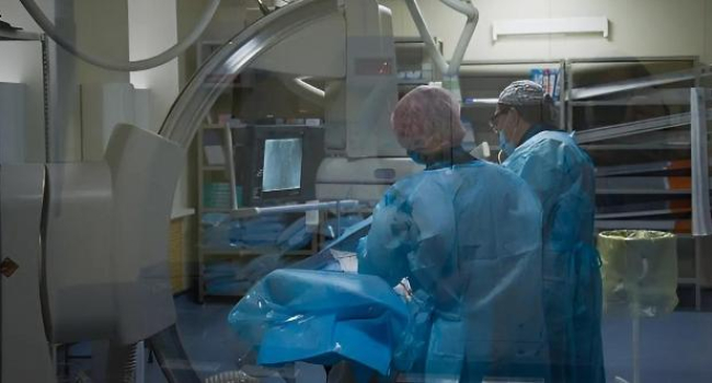
Scientists have introduced Histotripsy as a revolutionary method for treating tumors — and it looks almost like something from science fiction.
Instead of scalpels, lasers, or high-dose radiation, the technique uses ultrasound — but focused in such a way that thousands of microscopic bubbles form inside the tumor. These bubbles rapidly expand and collapse, literally breaking the tumor tissue into tiny particles. The results of the study were published in Science Translational Medicine.
The method is completely noninvasive: no incisions, no burns, and no radioactive exposure. Doctors can see everything on a screen in real time and precisely direct the “beam” at the tumor area, controlling the destruction locally so that healthy organs remain unharmed.
The first clinical trials have already produced encouraging results. Histotripsy has shown effectiveness in treating liver tumors: patients experienced fewer complications compared to traditional surgery and were even able to go home the same day.
Interestingly, the effect is not only local. When the tumor is destroyed, its fragments and “danger signal” molecules enter the bloodstream and can activate the immune system. This helps the body begin to recognize cancer cells on its own and fight them. In several experiments, immune responses were observed even against distant metastases, opening the door to combined approaches such as Histotripsy plus immunotherapy.
Researchers are also exploring the method for other applications: treating benign tumors, valve-related heart conditions, and even neurosurgery — thanks to its precision and gentle impact.
Another major advantage is that Histotripsy is suitable for patients who cannot undergo surgery or radiotherapy, allowing treatment with lower risk and a smoother recovery. It is possible that in the coming years, this technology will become a standard tool in oncology.