make preferable
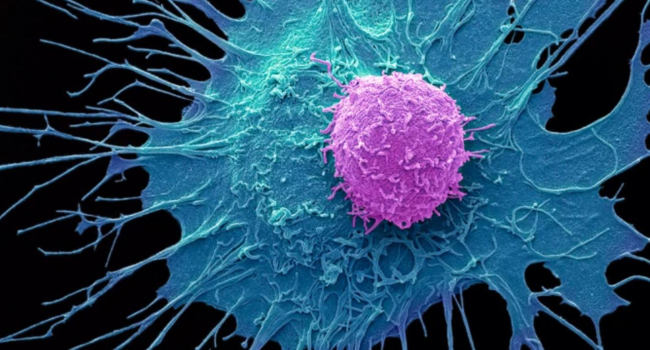
A new study shows how modern imaging techniques make it possible to literally “look inside” tumors and track the behavior of cancer cells with unprecedented precision.
The work was published in the journal Nature Methods.
Scientists have developed and applied new imaging tools that allow them to observe how individual cancer cells interact with their environment within a tumor. This is important because a tumor is not just a mass of identical cells, but a complex ecosystem in which cells can behave differently and even compete with one another.
Using these technologies, researchers were able to see how cells move, change their activity, and respond to external signals. It is especially significant that such methods make it possible to detect differences between cells within the same tumor—for example, which ones are more aggressive or resistant to treatment.
One of the key findings of the study is the significant heterogeneity of tumors. Even within a single tumor, different types of cells can coexist, varying in behavior and sensitivity to therapy. This partly explains why some treatments do not work for all patients or lose effectiveness over time.
The new imaging tools also help to better understand the mechanisms of metastasis—the process by which cancer cells spread throughout the body. By tracking cell movement, scientists gain a clearer picture of which cells are capable of leaving the primary tumor and forming secondary tumors.
In the future, such technologies could significantly influence cancer treatment. A more detailed understanding of cell behavior will enable the development of personalized therapies, allowing treatment to be tailored to the specific structure of a patient’s tumor.