


- Latest news▼
-
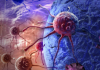
15:47, September 26 Scientists develop device to obtain live cells from suspicious tissue for early cancer detection
-
13:34, September 26 Virtual forest walk and classical music accelerated blood pressure reduction in emergency patients

-
11:05, September 26 Armenian pediatric oncologist Gevorg Tamamyan has been nominated by Botswana for the position of WHO Director-General

-
10:28, September 26 Could Earth’s magnetic field affect the aging process?

-
08:26, September 26 AI from Spain improves dermatological diagnosis

-
23:24, September 25 Study: Loneliness and social isolation may cost up to six years of healthy life

-
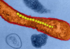
21:25, September 25 Magnetic bacteria extend worms’ lifespan by more than 43%, study finds
-
19:03, September 25 Chronic pain after cesarean delivery can affect women for years, study finds
-
16:58, September 25 Breath test may help distinguish cancerous lung nodules from benign ones, study finds

-
14:42, September 25 Study links overcrowding in emergency departments to hundreds of deaths each week
-
12:39, September 25 Experimental RNA therapy may help prevent muscle loss during GLP-1 weight loss

-
10:36, September 25 Introduction of solid foods triggers early functional changes in the infant gut microbiome

-
08:30, September 25 Lund University receives $19.4 million NIH grant to develop Alzheimer’s blood test
-
22:21, September 24 When Visual Signals Conflict, the Brain Works to Reach a Consensus

-
20:19, September 24 Coffee Can Disrupt Deep Sleep Even Without Keeping You Awake

All materials
Scientists develop device to obtain live cells from suspicious tissue for early cancer detection
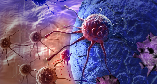
Scientists at the Massachusetts Institute of Technology (MIT) and Johns Hopkins University have developed a small microfluidic device that can extract living cells from precisely selected areas of tissue. In the future, the technology could help detect ovarian cancer at an earlier stage, study how tumors develop, and tailor treatment to individual patients.
The study was published in the journal Device.
One of the main challenges in ovarian cancer is that the disease is often diagnosed at a late stage. When detected early, the five-year survival rate can exceed 90%, while for patients diagnosed at stages III or IV, it is less than 50%.
About 20 years ago, scientists discovered that many cases of high-grade serous ovarian cancer, the most common type of the disease, likely originate in the fallopian tubes. However, detecting the earliest pathological changes in this area is difficult: precancerous lesions can be extremely small, and obtaining biological material from them for analysis is challenging.
The new device was developed by a team led by MIT mechanical engineering professor Kripa Varanasi, in collaboration with researchers at Johns Hopkins University. The 3D-printed device is placed directly on the surface of the tissue.
The device first creates a sealed contact with the tissue using a vacuum. Fluid is then passed through a small channel. The resulting flow produces controlled mechanical forces on the tissue surface, gently detaching living cells from a small area while leaving the surrounding tissue largely undisturbed.
The researchers tested the technology on human fallopian tubes that had been surgically removed. The extracted cells remained viable and multiplied significantly better in laboratory cultures than cells obtained using conventional methods. The researchers were also able to grow organoids from the cells — three-dimensional cellular models that can be used to study diseases and test drugs.
Conventional pathological diagnosis involves a different process. After tissue is removed, it is placed in a preservative solution, cut into thin sections, and examined under a microscope. This approach allows doctors to study the structure of the tissue in detail, but the cells no longer remain alive after processing and cannot be used for further culture.
According to the researchers, the ability to first identify a suspicious area and then obtain living cells from it could significantly expand the possibilities for diagnosis and research. The cells could be used to study the earliest changes associated with tumor development, create organoids, and test how tumor cells respond to different drugs.
The experiments also showed that the intensity of the fluid's mechanical action can be adjusted depending on the type of cells. According to the authors, this could potentially allow the technology to be applied not only to ovarian cancer but also to other types of tumors.
At the initial stage, the researchers plan to use the device on tissue that has already been surgically removed. This approach, they say, could simplify the subsequent medical regulatory process. In the future, the team hopes to adapt the device to obtain living cells directly from the body.
If the approach can eventually be used safely in patients, it could potentially combine targeted sampling with the preservation of viable cells, giving doctors and researchers additional information about the earliest stages of tumor development.
Если хотите, могу также сделать более близкий к оригиналу английский вариант, без стилистического рерайта.
Follow NEWS.am Medicine on Facebook and Twitter




- Most read
month
week
day
- Springer Nature: Scientists show that boys and girls perceive the world differently from the first hours of life 545
- Florida Records One of the Largest Dengue Outbreaks in Decades: One Person Has Died 539
- A&D: Residents of Two Amazonian Indigenous Communities Showed Unusually Low Rates of Dementia 482
- PLOS Medicine: More REM Sleep Is Associated With a Lower Risk of 83 Diseases 469
- GLP-1 Drugs Linked to Nutritional Deficiencies in Nearly 1 in 6 Children 464
- Lund University receives $19.4 million NIH grant to develop Alzheimer’s blood test 458
- Stopping GLP-1 drugs may quickly reduce their heart-protective benefits 451
- After 30 Years of Research, First Drug Developed for Rare Progressive Brain Disease 451
- JAMA: High Salivary Cortisol Levels Are Associated With Memory Decline 446
- F&F: Green oat extract may protect eyes from dryness caused by sleep deprivation 443
- Flu Vaccination Has Started in Armenia: Foreign Nationals Can Also Get Vaccinated Free of Charge 435
- NEG: 71% of People with Pulmonary Nodules Are Non-Smokers 434
- Rare Pregnancy Infections Linked to Higher Risk of Autism 431
- BMJ: Brisk Walking and Jogging Most Effective for Strengthening Bones 429
- Introduction of solid foods triggers early functional changes in the infant gut microbiome 420
- Find us on Facebook
- Poll