make preferable
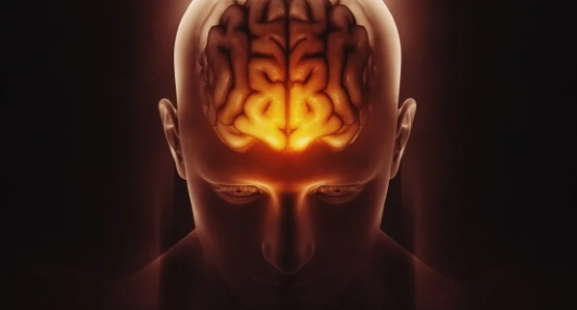
Scientists have discovered an unexpected effect: after a stroke, some areas of the brain may appear “younger” than expected for a person’s age. This may reflect the brain’s ability to reorganize and compensate for damage. The study was published in The Lancet Digital Health.
Researchers analyzed MRI scans of more than 500 post-stroke patients from 34 scientific centers across eight countries. Using artificial intelligence methods, they assessed the so-called “brain age”—an indicator reflecting the condition of brain tissue. The model analyzed 18 different brain regions and compared their biological age with the patient’s actual age.
The results were paradoxical. In the damaged hemisphere, a stroke accelerated tissue aging, while in the opposite hemisphere, a “rejuvenation” effect was observed.
“This may indicate that the brain is actively reorganizing itself, strengthening the function of undamaged networks,” explained study co-author Hosung Kim.
The effect was particularly pronounced in the frontoparietal network—the region responsible for movement planning, attention, and coordination. In patients with severe motor impairments, these areas showed a more “youthful” profile.
According to the researchers, this does not mean full recovery of function but rather points to compensatory mechanisms: the brain attempts to redistribute workload when damaged areas can no longer cope. A key role in the discovery was played by artificial intelligence—a graph neural network trained on tens of thousands of MRI scans. This approach made it possible to detect subtle changes that cannot be identified through standard analysis.
Project leader Arthur Toga noted that combining data from around the world helped reveal patterns that are not visible in smaller studies.
In the future, scientists plan to track patients at different stages of recovery. This could lead to the development of personalized rehabilitation programs that take into account individual patterns of brain reorganization.