make preferable
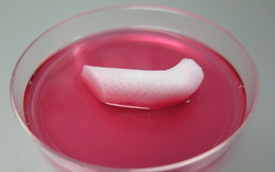
Rather than attempting to create a series of channels for blood to flow through, the scientists designed the printer to leave a series of micro-channels that allow nutrients and oxygen to flow through the growing tissue once implanted and develop a system of blood vessels on its own. If the technology can be perfected, the device could solve one of the biggest obstacles to creating 3D-printed organs: how to nourish masses of manufactured tissue.
He directs the Wake Forest Institute for Regenerative Medicine, in Winston-Salem, N.C.
The Wake Forest scientists who developed the new 3D printer obtained accurate 3D models of human body parts using CT and MRI scans-a procedure which could eventually be replicated to provide tailor-made organs to human patients.
But researchers in the United States report in Nature Biotechnology that they have harnessed a sophisticated, custom-designed 3D printer to print living muscle, cartilage and bone to fix battlefield injury.
They used bio-degradeable plastic to form the tissue's shape and water-based gels that contain cells, as well as a temporary outer structure to maintain shape of the tissue being printed. Atala and other researchers have developed methods to bioprint artificial skin and bladders.
Ear made with new 3D bioprinter.
Several teams of researchers, including the group at Wake Forest, have been working on techniques for growing thicker layers of tissue.
"We are also using similar strategies to print solid organs", he added. The bioprinter builds microchannels into the polymer scaffolding for the manufactured tissue.
Another limitation, Green said, has been the lack of blood vessels in bioprinted tissue: Larger structures are not possible without blood vessels to supply nutrients and oxygen.
And, to show that construction of a human-sized bone structure, jaw bone fragments were printed using human stem cells.
The process, which is thought not to cause any harm to the living cells, initially stuggled to ensure the engineered structures "lived" long enough to integrate into the body.
They then printed out an ear-shaped piece of cartilage, a muscle, and a piece of a jawbone, and implanted them in mice. Artificial muscle fibers were implanted in rats.
As 3D bioprinting moves forward, Green said, it will probably focus first on simpler structures that don't move or bear weight - including the ears, nose or bones in the skull - before trying to tackle more complicated tissue, or organs such as the heart, kidneys and pancreas.
While it has yet to be used in humans, Atala and his colleagues say that the bioprinter's principles are the same in people as they are in animals.
Atala said his team plans to implant bioprinted cartilage, bone and muscle tissue into patients in the future, with funding from the Armed Forces Institute of Regenerative Medicine.
In addition to Atala, the co-authors of the study in Nature Biotechnology, "A 3D Bioprinting System to Produce Human-Scale Tissue Constructs With Structural Integrity", include Hyun-Wook Kang, Sang Jin Lee, Carlos Kengla and James Yoo.