- Latest news▼
-
19:41, April 25 Children’s Hospital Los Angeles and International Center of Professional Development Allergy/Immunology Conference

-
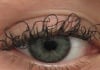
17:31, April 25 JAMA: patient grew long, curly eyelashes because of chemotherapy
-
11:08, April 25 Mpox epidemic declared in Republic of the Congo

-
08:31, April 25 OU: quitting smoking 4 times more likely to cure laryngeal cancer

-
01:20, April 25 Paralyzed man in China writes hieroglyphs using neural implants placed in his brain

-
15:11, April 24 Zombie deer disease possibly linked to hunters’ deaths

-
12:27, April 23 Appetite: Scientists found out the secret to the appeal of large portions of fast food

-
10:33, April 23 Scientists test new approach to fighting viruses

-
08:38, April 23 Ketamine may help with postpartum depression

-
22:12, April 22 Unhealthy amount of sugar found in baby food products of a well-known brand

-
19:41, April 22 Air pollution puts health of more than 1.6 billion workers globally at risk

-
17:25, April 22 Scientists found baked goods and lack of sleep to be more dangerous than alcohol

-
16:02, April 22 342 cases of measles recorded in Armenia so far in 2024

-
15:29, April 22 BrainStimulation: electrical brain stimulation alleviates anxiety and depression in the elderly

-
08:27, April 22 Cognitively stimulating jobs in midlife could lower dementia risk in old age, study finds

All materials
Brain scan shows a cyst squishing a man's brain against his skull
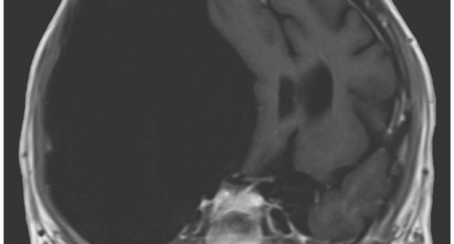
You see that massive black patch in this MRI scan? That’s a super-sized cyst lodged in between the brain and the skull.
The image was published online in the New England Journal of Medicine’s Image in Clinical Medicine this week.
This strange medical case recently came to light when a 27-year-old man was rushed to the emergency department at Metro Health Hospital in Wyoming after suffering from his first seizure. He told the doctors he had been suffering from frequent headaches and falls for over three years.
A prompt magnetic resonance imaging scan of his brain revealed the problem: a massive arachnoid cyst squishing the brain against the skull. The cyst is estimated to measure 12.3 by 16.5 by 7.9 centimeters (4.8 by 6.4 by 3.1 inches).
Arachnoid cysts are a thin sac filled with cerebrospinal fluid, a colorless fluid found in the brain and spinal cord. These specific cysts are found between the brain or spinal column and one of the membrane layers that surround the brain. The exact cause of them is unknown. Although, fear not, this is a particularly large one.
The doctors performed a craniotomy on the man. This is a procedure where a small portion of the skull is removed during surgery to allow them to drain the fluid and relieve the pressure. Follow-up imaging showed that this actually did not change the size of the cyst. The man still suffers from headaches every day and is fending off more seizures with anti-epileptic therapy.

Follow NEWS.am Medicine on Facebook and Twitter

- Video


- Event calendar
- Children’s Hospital Los Angeles and International Center of Professional Development Allergy/Immunology Conference
- First Armenian-German Conference entitled “Heart Failure Spring School”
- Allogeneic bone marrow transplant in case of hematological malignancy performed in Armenia for first time
All materials
- Archive
- Most read
month
week
day
- JAMA Oncology: Urine test can help rule out high-grade prostate cancer with almost 100% accuracy, study shows 1297
- Scientists grow human mini-lungs in lab 1175
- Next pandemic likely to be triggered by flu - scientists 946
- Scientists found baked goods and lack of sleep to be more dangerous than alcohol 925
- 342 cases of measles recorded in Armenia so far in 2024 879
- Scientists develop new method to safely stimulate immune cells to fight cancer 785
- Cognitively stimulating jobs in midlife could lower dementia risk in old age, study finds 781
- Blood test can determine who is at risk of developing multiple sclerosis - scientists 781
- BrainStimulation: electrical brain stimulation alleviates anxiety and depression in the elderly 721
- Ketamine may help with postpartum depression 662
- Unhealthy amount of sugar found in baby food products of a well-known brand 657
- Appetite: Scientists found out the secret to the appeal of large portions of fast food 653
- Air pollution puts health of more than 1.6 billion workers globally at risk 652
- Scientists test new approach to fighting viruses 637
- Zombie deer disease possibly linked to hunters’ deaths 546
- Find us on Facebook
- Poll